Visually stunning and easy to use, this volume in the highly regarded Diagnostic Pathology series covers the normal histology of every organ system. This edition incorporates the most recent scientific and technological knowledge in the field to prov ...view more
Visually stunning and easy to use, this volume in the highly regarded Diagnostic Pathology series covers the normal histology of every organ system. This edition incorporates the most recent scientific and technological knowledge in the field to provide a comprehensive overview of all areas of normal histology, including introductory chapters on electron microscopy, immunofluorescence, immunohistochemistry and histochemistry, the cell, and the basic organization of tissues. With nearly 1,800 outstanding images, this reference is an invaluable diagnostic aid for every practicing pathologist, resident, or fellow.
Visually stunning and easy to use, this volume in the highly regarded Diagnostic Pathology seriescovers the normal histology of every organ system. This edition incorporates the most recent scientific and technological knowledge in the field to provide a comprehensive overview of all areas of normal histology, including introductory chapters on electron microscopy, immunofluorescence, immunohistochemistry and histochemistry, the cell, and the basic organization of tissues. With nearly 1,800 outstanding images, this reference is an invaluable diagnostic aid for every practicing pathologist, resident, or fellow.
New to this edition
Thoroughly updated content throughout
, with all-new chapters on synovium and histologic artifacts, a thoroughly revised skeletal muscle chapter that now addresses normal histology in the setting of neuromuscular biopsy, and coverage of additional histologic variations that cause diagnostic confusion
New content on immunohistochemistry; more image examples of newly recognized normal variations, mimics, and pitfalls; and expanded text in many sections for greater clarity and ease of reference
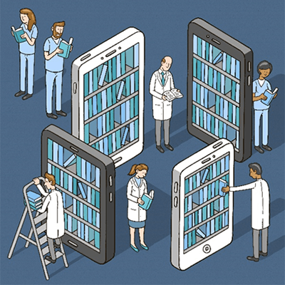
Expert Consult™ eBook version included with purchase. This enhanced eBook experience allows you to search all of the text, figures, and references from the book on a variety of devices.
Key Features
Unparalleled visual coverage
with carefully annotated photomicrographs, spectacular gross images, electron micrographs, and medical illustrations
Time-saving reference features
include bulleted text, a variety of test data tables, key facts in each chapter, annotated images, and an extensive index
Author Information
By Matthew R. Lindberg, MD, Director, Soft Tissue Pathology Division, Associate Professor of Pathology, University of Arkansas for Medical Sciences, Little Rock, Arkansas
Bring it with you - The Bookshelf app allows you to access books on your laptop, tablet or mobile, so your ebooks go where ever you are - online or offline.
Take notes - Highlight, bookmark and take notes and highlights automatically stay in sync no matter where you make them.
Listen to eBooks - When you need to go screenless, the Text-to-speech tool will read your book aloud.
Powerful search - The searching capabilities allow you to search keywords through all your eBooks, the entire Bookshelf Library and well as on Wikipedia.
Ecological - Manage your environmental impact with paperless books.
How to read your VitalSource eBooks
Purchase your VitalSource eBook on the Elsevier Health site
https://www.eu.elsevierhealth.com/diagnostic-pathology-normal-histology-9780323548045.html101026Diagnostic Pathology: Normal Histologyhttps://www.eu.elsevierhealth.com/media/catalog/product/9/7/9780323548045_9.jpg168.14176.99EURInStock/Medicine and Surgery/Pathology/eBooks983752550401745498285145120<p>Visually stunning and easy to use, this volume in the highly regarded <i>Diagnostic Pathology</i> series<b> </b>covers the normal histology of every organ system. This edition incorporates the most recent scientific and technological knowledge in the field to provide a comprehensive overview of all areas of normal histology, including introductory chapters on electron microscopy, immunofluorescence, immunohistochemistry and histochemistry, the cell, and the basic organization of tissues. With <b>nearly 1,800 outstanding images</b>, this reference is <b>an invaluable diagnostic aid</b> for every practicing pathologist, resident, or fellow.</p> <p>Visually stunning and easy to use, this volume in the highly regarded <i>Diagnostic Pathology</i> series<b> </b>covers the normal histology of every organ system. This edition incorporates the most recent scientific and technological knowledge in the field to provide a comprehensive overview of all areas of normal histology, including introductory chapters on electron microscopy, immunofluorescence, immunohistochemistry and histochemistry, the cell, and the basic organization of tissues. With <b>nearly 1,800 outstanding images</b>, this reference is <b>an invaluable diagnostic aid</b> for every practicing pathologist, resident, or fellow.</p>00add-to-cart97803235480452017ProfessionalBy Matthew R. Lindberg, MD20182E-BookOther1800 illustrations (1800 in full color)Elsevier5008 Nov 2017IN STOCKBy <STRONG>Matthew R. Lindberg</STRONG>, MD, Director, Soft Tissue Pathology Division, Associate Professor of Pathology, University of Arkansas for Medical Sciences, Little Rock, ArkansaseBooksDiagnostic PathologyNoNoNoNoPlease SelectPlease SelectPlease Select